Medical physics
🎯What you need to be able to do
- Explain the principles of the generation and detection of ultrasound using a piezoelectric transducer.
- Explain the main principles behind the use of ultrasound to obtain diagnostic information about internal structures.
- Define specific acoustic impedance, \( Z = \rho c \), and use the intensity reflection coefficient at a boundary.
- Recall and use \( I = I_0e^{-\mu x} \) for the attenuation of ultrasound in matter.
- Explain the principles of the production of X-rays by electron bombardment of a metal target, and use \( hc/\lambda_{\min} = eV \) for the minimum wavelength produced.
- Understand the use of X-rays in imaging, and what is meant by the contrast of an image.
- Recall and use \( I = I_0e^{-\mu x} \) for the attenuation of X-rays in matter.
- Understand the purpose of computed tomography and how a CT scan builds a three-dimensional image from many slices.
- Explain the principles of positron emission tomography, including annihilation and the detection of coincident gamma photons.
📚The physics
Ultrasound. A piezoelectric crystal — usually lead zirconate titanate — changes shape when a p.d. is applied across it and produces a p.d. when it is deformed. The same crystal therefore both transmits pulses and detects the echoes. It is driven at its resonant frequency, typically 1–15 MHz. Depth is calculated from the time delay: \( d = ct/2 \), and that factor of two is there because the pulse travels there and back.
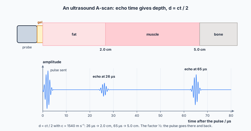
Impedance and the gel. The specific acoustic impedance is \( Z = \rho c \). At a boundary the fraction of intensity reflected is \( I_r/I_0 = (Z_2 - Z_1)^{2}/(Z_2 + Z_1)^{2} \). Air and soft tissue have wildly different impedances, so a thin air gap between probe and skin reflects almost all of the beam and nothing reaches the patient. The gel has an impedance close to that of skin, which is why it is not merely a lubricant.
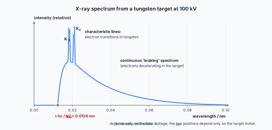
X-ray production. Electrons are accelerated through tens of kilovolts onto a metal target. Most of their energy becomes internal energy in the target — which is why the anode rotates and is cooled — and a small fraction becomes X-rays. The continuous braking spectrum has a sharp minimum wavelength set by the accelerating voltage, \( hc/\lambda_{\min} = eV \), corresponding to an electron that loses all its energy in one interaction. Superimposed on it are characteristic line peaks from electron transitions in the target atoms.
Attenuation. \( I = I_0e^{-\mu x} \). The attenuation coefficient \(\mu\) depends strongly on the atomic number of the material, which is why bone stands out against soft tissue and why barium and iodine work as contrast media. Contrast is the difference in attenuation between adjacent structures — it is what lets you tell one tissue from another on the image. (Sharpness, which depends on the geometry of the source and detector rather than on attenuation, is a separate matter and is not part of this syllabus.)
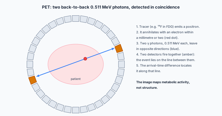
PET. A positron-emitting tracer such as fluorine-18 in fluorodeoxyglucose is given to the patient. Each emitted positron annihilates with a nearby electron within a millimetre or two, producing two gamma photons of 0.511 MeV each travelling in opposite directions. A ring of detectors records pairs arriving in coincidence; the small difference in arrival times locates the annihilation along the line joining the two detectors. The result is a map of metabolic activity rather than of structure, which is what makes PET complementary to CT rather than a substitute for it.
✏️Worked example
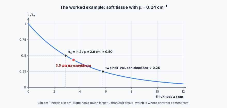
\( I/I_0 = e^{-\mu x} = e^{-0.24 \times 3.5} = e^{-0.84} = 0.43 \), so 43% is transmitted.
🔭See it happen
Most schools cannot run an X-ray set, but an ultrasound distance sensor from a robotics kit does the physics honestly: it emits a pulse, times the echo and halves it. Have students measure a known distance with it and then work backwards to the speed of sound in air. For attenuation, a gamma source, a set of lead absorbers and a Geiger counter give the same exponential law in a form you can plot.
🔗Go deeper — other people’s work
The links below are not mine. They are here because they are good, and they may move or disappear without warning.
- The British Institute of Radiology and the IAEA both publish accessible introductions to diagnostic imaging physics.
- HyperPhysics, “Medical Imaging”.