Cells and microscopy
🎯What you need to be able to do
- State the cell theory.
- Identify the parts of animal, plant and bacterial cells and describe their functions.
- Compare prokaryotic and eukaryotic cells.
- Explain how specialized cells are adapted to their functions, and what stem cells are.
- Describe the levels of organization: cell, tissue, organ, organ system, organism.
- Use a light microscope and calculate magnification and actual size.
🔬The cell theory
The cell theory states that: all living things are made of one or more cells; the cell is the smallest unit of life; and all cells come from pre-existing cells, by division. Robert Hooke named “cells” in 1665 after looking at cork; Antonie van Leeuwenhoek saw living single-celled organisms with his own lenses a few years later. Better microscopes, especially the electron microscope, revealed the structures inside cells.
🦠Animal and plant cells
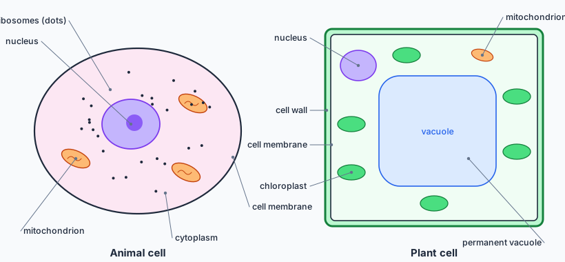
| Structure | Function | Found in |
|---|---|---|
| nucleus | contains DNA in chromosomes; controls the cell’s activities | plants and animals |
| cytoplasm | jelly where most chemical reactions happen | all cells |
| cell membrane | controls what enters and leaves the cell (partially permeable) | all cells |
| mitochondria | site of aerobic respiration, releasing energy | plants and animals |
| ribosomes | make proteins | all cells |
| cell wall | made of cellulose; supports the cell and keeps its shape | plants (and fungi, bacteria, with other materials) |
| chloroplasts | contain chlorophyll; absorb light for photosynthesis | plant cells in green parts |
| permanent vacuole | contains cell sap; keeps the cell firm (turgid) | plants |
🦟Prokaryotes and eukaryotes
Eukaryotic cells (animals, plants, fungi, protoctists) have a nucleus and membrane-bound organelles such as mitochondria. Prokaryotic cells (bacteria) are much smaller and simpler: no nucleus — their DNA is a single loop in the cytoplasm, often with extra small rings called plasmids — and no mitochondria or chloroplasts. They have a cell wall (not cellulose), ribosomes, and sometimes a flagellum for movement.
🦺Specialized cells and stem cells
In a multicellular organism, cells differentiate: they become specialized, with a structure suited to one job.
- Red blood cell — no nucleus, packed with haemoglobin, biconcave shape for a large surface area: carries oxygen.
- Nerve cell (neurone) — long axon and many branched endings: carries electrical impulses over long distances.
- Sperm cell — tail for swimming, many mitochondria for energy, enzymes in the head to penetrate the egg.
- Root hair cell — long extension that gives a huge surface area for absorbing water and mineral ions; no chloroplasts.
- Palisade cell — packed with chloroplasts near the top of the leaf: maximum photosynthesis.
- Ciliated cell — hair-like cilia sweep mucus and trapped dust out of the airways.
Stem cells are unspecialized cells that can divide and become many types of cell. Embryonic stem cells can become almost any cell; adult stem cells (in bone marrow, for example) form a limited range. Plants keep stem cells in meristems at their tips all their lives. Stem cells could be used to treat diseases such as leukaemia and diabetes, which raises ethical questions about the use of embryos (Topic 16).
🧱Levels of organization
Cells of the same type form a tissue (muscle tissue); several tissues form an organ (the stomach has muscle, glandular and lining tissue); organs working together form an organ system (the digestive system); organ systems make up the organism.
🔍Microscopes and magnification
A light microscope can magnify up to about ×1500 and shows cells and the nucleus; an electron microscope magnifies over ×1 000 000 with far higher resolution (the ability to see two close points as separate), revealing mitochondria and ribosomes in detail. To use a light microscope: start with the lowest-power objective, focus with the coarse knob, then switch to higher power and use only the fine knob.
✏️Worked example: how big is the cell really?
1. Rearrange. actual size = image size ÷ magnification = 45 mm ÷ 750 = 0.060 mm.
2. Convert. 0.060 mm × 1000 = 60 µm.
🌎Science in context: seeing smaller
Each advance in microscopy changed biology: light microscopes revealed cells and bacteria, the electron microscope revealed viruses and organelles, and cryo-electron microscopy (Nobel Prize 2017) now shows the shapes of individual protein molecules — which helped scientists design COVID-19 vaccines quickly. Technology and scientific understanding advance together, a useful point for any Criterion D discussion of how science develops.
🧠Quick check
1. Name three structures found in plant cells but not animal cells.
Cell wall, chloroplasts, large permanent vacuole.
2. What is the function of mitochondria?
They are the site of aerobic respiration, which releases energy for the cell.
3. Give two differences between a bacterial cell and an animal cell.
Bacteria have no nucleus (DNA loop and plasmids in the cytoplasm), no mitochondria, and do have a cell wall; they are also much smaller.
4. How is a root hair cell adapted to its function?
Its long projection gives a large surface area for absorbing water and mineral ions; it has no chloroplasts because it is underground.
5. Put in order from smallest to largest: organ, cell, organ system, tissue.
Cell, tissue, organ, organ system.
6. An eyepiece is ×10 and the objective ×40. A cell appears 20 mm wide. What is its actual width?
Magnification = ×400. Actual size = 20 ÷ 400 = 0.05 mm = 50 µm.
📝Worksheet
Test yourself on the whole topic with a printable worksheet: questions for all four criteria, from recall to a design task, a data-analysis question and a short reflection, with a full mark scheme.
Worksheets are for members — sign in or join. The topic 1 worksheet is a free sample.