Analytical techniques
🎯What you need to be able to do
- Describe thin-layer chromatography: stationary phase, mobile phase, Rf, solvent front and baseline; interpret and explain Rf values.
- Describe gas–liquid chromatography: stationary and mobile phases, retention time; interpret chromatograms for percentage composition; explain retention times.
- Interpret carbon-13 NMR spectra: carbon environments and possible structures; predict the number of peaks.
- Interpret proton NMR spectra: environments from chemical shift, relative numbers from peak areas, neighbours from splitting (n + 1 rule), and possible structures; predict shifts and splitting.
- Describe the use of TMS, deuterated solvents such as CDCl3, and D2O exchange to identify O–H and N–H protons.
📚The chemistry
AS analysis (topic 22) found functional groups (infrared) and molecular mass (mass spectrometry). This topic adds ways to separate mixtures (chromatography) and to map the carbon and hydrogen skeleton of a molecule (NMR). All chromatography works on one principle: components of a mixture are distributed between a stationary phase and a mobile phase, and those that interact more with the stationary phase move more slowly.
37.1 Thin-layer chromatography (TLC)
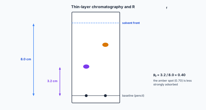
- Stationary phase: a thin layer of a polar solid such as aluminium oxide (or silica) on a solid support (a glass or plastic plate).
- Mobile phase: a solvent, polar or non-polar, which moves up the plate by capillary action.
- Baseline: the pencil line where the samples are spotted, above the solvent level so they do not simply dissolve away.
- Solvent front: how far the solvent has travelled when the plate is removed; mark it immediately.
Both distances are measured from the baseline. Rf is between 0 and 1 and has no units. Under the same conditions (same stationary phase, solvent and temperature) a substance always has the same Rf, so comparing with standards identifies components. Colourless spots are shown with UV light or a locating agent.
Explaining Rf values: a component that is strongly adsorbed onto the stationary phase (for polar aluminium oxide, a polar molecule, especially one that can hydrogen bond) moves slowly: low Rf. A component that is more soluble in the mobile phase and interacts less with the stationary phase moves further: high Rf.
37.2 Gas–liquid chromatography (GLC)
- Stationary phase: a high-boiling-point, non-polar liquid coated on a solid support, packed in or lining a long, thin, coiled column in an oven.
- Mobile phase: an unreactive carrier gas such as helium or nitrogen.
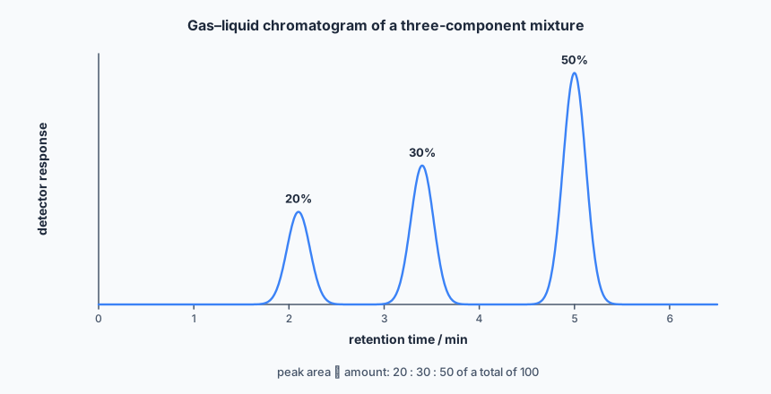
- Retention time: the time from injection to the component reaching the detector. Each component, at a fixed temperature and flow rate, has a characteristic retention time.
Explaining retention times: a component that is more soluble in the stationary liquid (interacts more strongly with it) is held back longer: longer retention time. With a non-polar stationary phase, larger, less volatile molecules with stronger id-id interactions are retained longest; volatile, small molecules emerge first.
Percentage composition: the area under each peak is proportional to the amount of that component. Percentage of a component = its peak area ÷ total of all peak areas × 100. (Where peaks are triangles, area ≈ ½ × base × height.)
37.3 Carbon-13 NMR
Each chemically different carbon environment gives one peak. Carbons are equivalent if they are in identical surroundings — related by the symmetry of the molecule. The position of the peak, its chemical shift δ (ppm), shows the type of environment; the data section gives ranges, for example sp3 alkyl 0–50, next to oxygen 50–70, alkene or arene 110–160, carboxyl or ester C=O 160–185, aldehyde or ketone C=O 190–220.
In 13C spectra (as usually recorded) peak height is not a reliable guide to the number of carbons, and there is no splitting to interpret: just count the peaks and read their shifts.
37.4 Proton (1H) NMR
A proton NMR spectrum gives four pieces of information:
- Number of peaks — the number of different proton environments.
- Chemical shift, δ — the type of environment, from the data section ranges: alkyl 0.9–1.7; alkyl next to C=O 2.2–3.0; next to an electronegative atom (CH2–O, CH2–Cl) 3.2–4.0; on an alkene 4.5–6.0; on an aromatic ring 6.0–9.0; aldehyde 9.3–10.5; carboxylic acid 9.0–13.0. Electronegative atoms nearby move a peak to higher δ.
- Relative peak area (integration) — the relative number of protons in each environment.
- Splitting pattern — the number of protons on the adjacent carbon atom(s). By the n + 1 rule, a peak is split into n + 1 lines by n equivalent protons on neighbouring carbons: singlet (no neighbouring H), doublet (1), triplet (2), quartet (3), multiplet (more, or several different sets).
Protons on the same carbon, or equivalent protons, do not split each other. The classic ethyl pattern is a triplet (3H) and a quartet (2H): the CH3 has two neighbours (CH2) so it is a triplet; the CH2 has three neighbours (CH3) so it is a quartet.
TMS, solvents and D2O
- Tetramethylsilane, TMS, Si(CH3)4, is the standard: its peak defines δ = 0. It is used because all 12 protons are equivalent (a single sharp peak), it absorbs upfield of almost all organic protons, it is inert and volatile (easily removed from the sample afterwards).
- Deuterated solvents, such as CDCl3, are used because deuterium (2H) does not give a signal in a proton spectrum, so the solvent does not swamp the sample’s peaks.
- D2O exchange: O–H and N–H protons have variable, sometimes broad peaks that are hard to assign. Shake the sample with D2O: these protons exchange with deuterium (R–OH + D2O ⇌ R–OD + HOD), and their peak disappears from a new spectrum. That identifies it. O–H protons usually appear as singlets, because rapid exchange stops them splitting or being split.
✏️Worked example
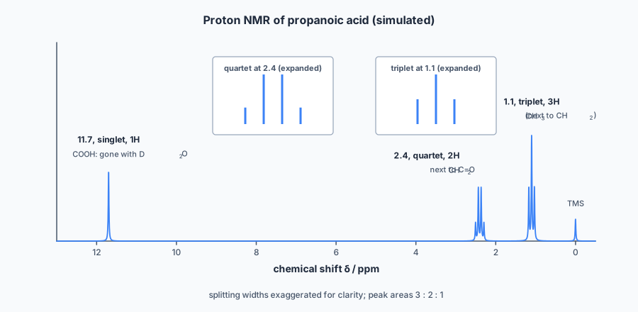
• δ 1.1, triplet, relative area 3
• δ 2.4, quartet, relative area 2
• δ 11.7, singlet, relative area 1; this peak disappears when D2O is added.
(a) Identify X, explaining each peak. (b) How many peaks would its 13C spectrum show, and in which range would the peak for the C=O carbon appear? (c) Methyl ethanoate is an isomer of X. Predict its proton NMR spectrum (number of peaks, their areas and splitting).
(a)
- δ 11.7, singlet, 1H, removed by D2O: an O–H proton; the shift is in the carboxylic acid range (9.0–13.0), so this is –COOH.
- δ 2.4, quartet, 2H: a CH2 next to C=O (range 2.2–3.0), split into a quartet by three neighbouring H, a CH3.
- δ 1.1, triplet, 3H: a CH3 in an alkyl environment (0.9–1.7), split into a triplet by two neighbouring H, the CH2.
Together: CH3–CH2–COOH. X is propanoic acid, which fits C3H6O2, and the areas add to 6 H.
(b) Three carbons, all in different environments: 3 peaks. The carboxyl C=O appears in the 160–185 range.
(c) Methyl ethanoate, CH3COOCH3, has two CH3 groups in different environments, and neither has H on the neighbouring atom (C=O on one side, O on the other): two singlets, each area 3. The CH3 attached to O is at the higher shift, in the 3.2–4.0 range.
📝Practise
Work through these, then reveal the answer. Each question targets a different objective from the list above.
1. On a TLC plate the solvent front is 8.0 cm from the baseline and a spot is 3.2 cm from the baseline. Calculate Rf, and explain what a lower Rf would mean on an aluminium oxide plate.
2. A gas chromatogram of a mixture shows three peaks with areas 24, 36 and 60 units. Calculate the percentage composition.
3. Predict the number of peaks in the 13C NMR spectra of (a) butanone; (b) 2-methylpropan-2-ol; (c) 1,4-dimethylbenzene.
4. Predict the proton NMR spectrum of ethanol, CH3CH2OH, in CDCl3, and what changes on adding D2O.
5. Give three reasons why TMS is used as the reference in NMR.
6. A compound C3H6O shows only one peak in its proton spectrum: a singlet at δ 2.2. Identify it and explain.
🔗Go deeper — other people’s work
These are external resources, not mine. If one stops working, tell me and everything above it on this page still stands.
- Chemguide (Jim Clark) — the NMR and chromatography pages
- SDBS (Spectral Database for Organic Compounds, AIST Japan) — real 1H and 13C spectra for thousands of compounds, for practice