Home › Learning Hub › IGCSE Biology › 11 Gas exchange in humans
Topic 11 · 11.1

Gas exchange in humans

Core and Extended · Papers 1–6

🎯What you need to be able to do

  • Describe the features of gas exchange surfaces and identify the parts of the breathing system.
  • Investigate and describe the differences between inspired and expired air, and the effect of physical activity on breathing.
  • Identify the internal and external intercostal muscles, state the function of cartilage, and explain ventilation EXTENDED.
  • Explain the composition of expired air, the link between exercise and breathing, and the roles of goblet cells, mucus and cilia EXTENDED.

📚The biology

The breathing system
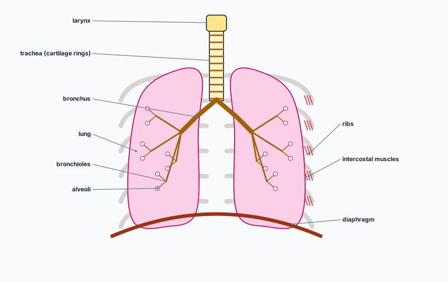
The human breathing system. The larynx at the top of the trachea, which has rings of cartilage. The trachea divides into two bronchi, which branch into bronchioles ending in tiny air sacs, the alveoli, inside the two lungs. Ribs surround the lungs, with intercostal muscles between them, and the domed diaphragm lies underneath.
Air passes larynx → trachea → bronchi → bronchioles → alveoli.

EXTENDED Cartilage in the trachea holds it open, so it does not collapse when air pressure inside falls.

Gas exchange surfaces
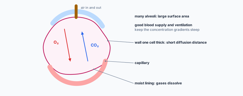
An alveolus, an air sac, wrapped by a capillary. Oxygen diffuses from the air in the alveolus into the blood; carbon dioxide diffuses from the blood into the alveolus. Labels: wall one cell thick, a short diffusion distance; moist lining, where gases dissolve; many alveoli give a large surface area; a good blood supply and ventilation keep the concentration gradients steep.
The four features of a gas exchange surface: large surface area, thin surface, good blood supply, good ventilation with air.

Inspired and expired air

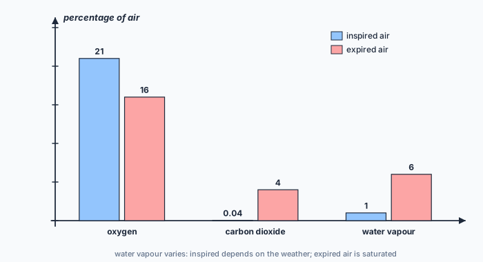
A bar chart comparing inspired and expired air. Oxygen: 21 percent in inspired air, 16 percent in expired air. Carbon dioxide: 0.04 percent inspired, 4 percent expired. Water vapour: about 1 percent inspired (it varies), 6 percent expired, which is saturated.
Expired air has less oxygen and more carbon dioxide and water vapour. (Typical values.)
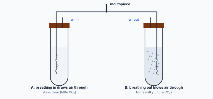
Two boiling tubes of limewater joined to one mouthpiece. Breathing in draws air through tube A, which stays clear; breathing out blows air through tube B, which turns milky, showing that expired air contains more carbon dioxide.
Breathe gently in and out through the mouthpiece; compare how long each tube takes to turn milky.

EXTENDED Expired air has less oxygen because oxygen diffuses into the blood and is used in respiration; more carbon dioxide because carbon dioxide produced in respiration diffuses into the alveoli; and more water vapour because water evaporates from the moist lining of the alveoli.

During physical activity both the rate and the depth of breathing increase. EXTENDED Muscles respire faster, so the carbon dioxide concentration in the blood rises; this is detected by the brain, which increases the rate and depth of breathing to remove carbon dioxide and take in more oxygen.

Ventilation EXTENDED
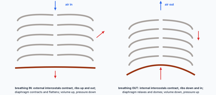
Two diagrams of the ribcage and diaphragm. Breathing in: the ribs are raised up and out and the diaphragm is flattened; air flows in. Breathing out: the ribs are lower and the diaphragm is domed upwards; air flows out.
EXTENDED Air always moves from higher to lower pressure: a bigger thorax means lower pressure inside, so air rushes in.
  • EXTENDED Breathing in: the external intercostal muscles contract, moving the ribs up and out; the diaphragm contracts and flattens. The volume of the thorax increases, the pressure falls below atmospheric pressure, and air is drawn in.
  • Breathing out: the external intercostals relax and the internal intercostal muscles contract (in forced breathing), moving the ribs down and in; the diaphragm relaxes and domes upwards. Volume decreases, pressure increases, and air is pushed out.

Protecting the airways EXTENDED
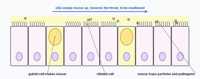
The lining of the trachea: a row of cells, mostly ciliated cells with hair-like cilia on top, with two goblet cells full of mucus. A layer of mucus above traps particles and pathogens, and the cilia sweep it up towards the throat to be swallowed.
EXTENDED Smoking damages the cilia, so mucus collects and smokers cough to clear it.

EXTENDED Goblet cells secrete mucus, which traps pathogens and particles. Ciliated cells have cilia that beat to sweep the mucus up to the throat, where it is swallowed, so pathogens do not reach the alveoli.

✏️Worked example

(a) Use the bar chart to calculate how many times more carbon dioxide there is in expired air than in inspired air. [1] (b) EXTENDED Explain the difference in oxygen between inspired and expired air. [2] (c) EXTENDED Describe how the diaphragm and external intercostal muscles cause air to enter the lungs. [4]

(a) \( 4 \div 0.04 = 100 \) times.

(b) Oxygen diffuses from the alveoli into the blood and is carried to cells, where it is used in aerobic respiration, so expired air contains less (16% compared with 21%).

(c) The external intercostal muscles contract, moving the ribs up and out; the diaphragm contracts and flattens. The volume of the thorax increases, so the pressure inside falls below atmospheric pressure, and air moves into the lungs.

Check it. Expired air still contains 16% oxygen, which is why rescue breaths in first aid can keep someone alive.
“The lungs expand and suck air in.” The lungs have no muscle. Muscles change the volume of the thorax; the pressure change moves the air.

📝Practise

In the style of the multiple-choice, theory and practical papers. EXTENDED marks Supplement content.

1. (Multiple choice.) Which is NOT a feature of a gas exchange surface? A: large surface area. B: thin surface. C: good blood supply. D: thick muscular wall.
D.
2. (Theory.) Name the structures, in order, that air passes through from the larynx to the alveoli. [2]
Trachea, bronchus (bronchi), bronchioles, alveoli.
3. (Practical.) In the limewater experiment, state the result for each tube and what it shows. [2]
The tube receiving expired air turns milky (sooner); the other stays clear (or changes much more slowly). Expired air contains more carbon dioxide than inspired air.
4. (Practical.) Describe how to investigate the effect of exercise on the rate of breathing. [3]
Count breaths per minute at rest; do a set exercise (e.g. step-ups for 2 minutes); count breaths per minute immediately after, and every minute until it returns to resting; repeat and take means.
5. (Theory.) EXTENDED State the function of the cartilage rings in the trachea. [1]
They keep the trachea open, so it does not collapse when air pressure falls.
6. (Theory.) EXTENDED Explain why expired air contains more water vapour than inspired air. [1]
Water evaporates from the moist lining of the alveoli into the air.
7. (Theory.) EXTENDED Explain why breathing becomes faster and deeper during exercise. [3]
Muscles respire faster and produce more carbon dioxide; the carbon dioxide concentration in the blood rises; this is detected by the brain, which increases the rate and depth of breathing.
8. (Theory.) EXTENDED Chemicals in tobacco smoke stop cilia beating. Explain why smokers are more likely to get lung infections. [3]
Mucus that traps pathogens is not swept up out of the airways; it collects in the lungs, so pathogens stay in the breathing system and can cause infection.

🔗Go deeper — other people’s work

These are external resources, not mine. If one stops working, tell me and everything above it on this page still stands.

  • British Lung Foundation — how your lungs work
  • Royal Society of Biology — the bell-jar model of the thorax