Blood vessels and blood
🎯What you need to be able to do
- Describe the structure of arteries, veins and capillaries; state the functions of capillaries; explain how structure relates to pressure and function EXTENDED.
- Identify the main blood vessels to and from the heart, lungs and kidneys; and the liver EXTENDED.
- List the components of blood, identify red and white blood cells, and state their functions; identify lymphocytes and phagocytes EXTENDED.
- State the roles of clotting; describe it as fibrinogen converted to fibrin EXTENDED.
📚The biology
Arteries, veins and capillaries
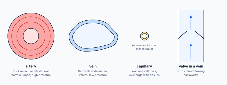
Capillaries exchange substances with the tissues: oxygen and nutrients pass out to the cells; carbon dioxide and other wastes pass in.
- EXTENDED Arteries carry blood at high pressure: their thick walls withstand it, the muscle and elastic tissue stretch and recoil to keep the blood flowing (you feel this as the pulse), and the narrow lumen maintains the pressure.
- Veins carry blood at low pressure: thin walls are enough, the wide lumen reduces resistance to flow, and valves prevent backflow.
- Capillaries: the wall is one cell thick, so the diffusion distance is short; they are narrow and form huge networks, so every cell is close to one; their walls are leaky, letting some plasma out to bathe the cells.
The main blood vessels
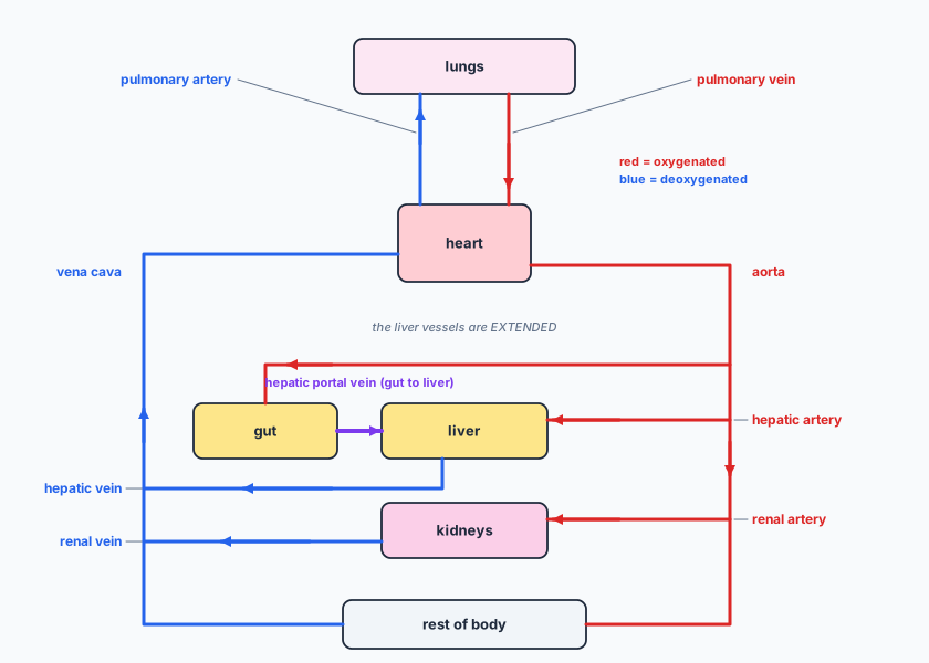
The pulmonary artery is the only artery carrying deoxygenated blood, and the pulmonary vein the only vein carrying oxygenated blood. EXTENDED The hepatic portal vein carries blood rich in absorbed nutrients from the gut straight to the liver.
Blood
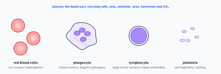
EXTENDED Lymphocytes produce antibodies; phagocytes engulf pathogens by phagocytosis.
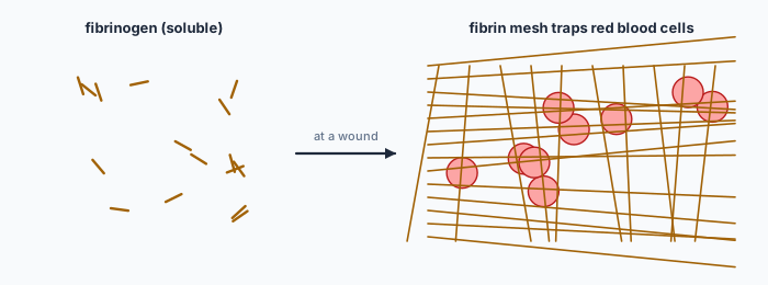
Blood clotting prevents blood loss and stops pathogens entering through a wound. EXTENDED At a wound, the soluble protein fibrinogen is converted to insoluble fibrin, which forms a mesh that traps red blood cells and forms a clot.
✏️Worked example
(a) A vein; low pressure.
(b) (i) Renal vein. (ii) Pulmonary artery.
(c) Their wall is one cell thick, so substances diffuse a short distance; they form large networks, giving a large surface area and bringing every cell close to a capillary; they are leaky, letting plasma out to supply the cells.
📝Practise
In the style of the multiple-choice and theory papers. EXTENDED marks Supplement content.
1. (Multiple choice.) Which component of blood carries urea to the kidneys? A: red blood cells. B: white blood cells. C: platelets. D: plasma.
2. (Theory.) State two differences in structure between an artery and a vein. [2]
3. (Theory.) State the function of haemoglobin, and name the cells that contain it. [2]
4. (Theory.) State two roles of blood clotting. [2]
5. (Theory.) Name the vessel that (a) brings oxygenated blood from the lungs to the heart, (b) carries blood to the kidneys. [2]
6. (Theory.) EXTENDED Explain why arteries have thick, elastic walls. [2]
7. (Theory.) EXTENDED Describe how a phagocyte and a lymphocyte can be distinguished in a blood smear, and state the function of each. [4]
8. (Theory.) EXTENDED Name the blood vessel that carries absorbed glucose from the small intestine to the liver. [1]
🔗Go deeper — other people’s work
These are external resources, not mine. If one stops working, tell me and everything above it on this page still stands.
- Royal Society of Biology — examining blood smears under the microscope
- NHS Blood and Transplant — what blood is made of