Home › Learning Hub › IGCSE Biology › 14a Nervous system and the eye
Topic 14 · 14.1–14.2

The nervous system and the eye

Core and Extended · Papers 1–6

🎯What you need to be able to do

  • Describe the CNS and PNS, the role of the nervous system, and identify sensory, relay and motor neurones.
  • Describe a reflex arc and a reflex action; describe a synapse and the events at it EXTENDED.
  • Describe sense organs; identify the parts of the eye and state their functions; explain the pupil reflex.
  • Explain the pupil reflex with antagonistic muscles, accommodation, and the roles of rods, cones and the fovea EXTENDED.

📚The biology

The nervous system

Electrical impulses travel along neurones. The nervous system coordinates and regulates body functions.
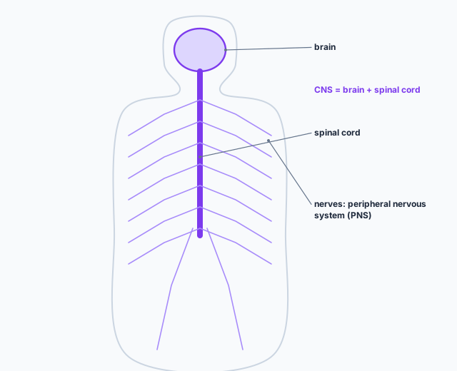
An outline of a human body showing the brain and spinal cord, which form the central nervous system, and nerves branching out to the rest of the body, which form the peripheral nervous system.
The central nervous system (CNS) is the brain and spinal cord; the peripheral nervous system (PNS) is the nerves outside them.
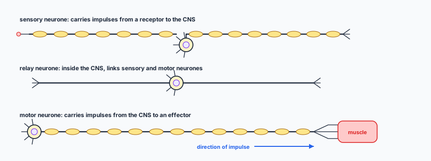
Three types of neurone. A sensory neurone runs from a receptor, with its cell body part-way along, to the CNS. A relay neurone, short and inside the CNS, links sensory and motor neurones. A motor neurone has its cell body in the CNS and a long axon ending on a muscle. An arrow shows the direction of the impulse.
Tell them apart by the position of the cell body: in the middle (sensory), in the CNS at one end (motor), or in a short neurone inside the CNS (relay).

Reflexes

A reflex action is a means of automatically and rapidly integrating and coordinating stimuli with the responses of effectors (muscles and glands). It needs no conscious thought, which saves time and protects the body.
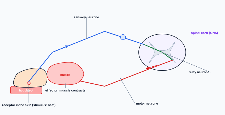
A reflex arc. A hand touches a hot object; a receptor in the skin is stimulated. A sensory neurone carries the impulse to the spinal cord, where a relay neurone passes it to a motor neurone. The motor neurone carries it to a muscle, the effector, which contracts and pulls the hand away.
Receptor → sensory neurone → relay neurone → motor neurone → effector.

A synapse is a junction between two neurones.
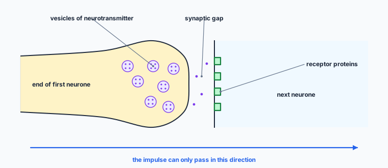
A synapse. The end of the first neurone contains vesicles of neurotransmitter. A narrow synaptic gap separates it from the next neurone, whose membrane carries receptor proteins. Neurotransmitter molecules cross the gap. An arrow shows the impulse passing in one direction only.
EXTENDED Only the first neurone has vesicles and only the second has receptors, so impulses cross in one direction.
  1. EXTENDED An impulse stimulates the release of neurotransmitter molecules from vesicles into the synaptic gap.
  2. The neurotransmitter molecules diffuse across the gap.
  3. They bind with receptor proteins on the next neurone.
  4. An impulse is then stimulated in the next neurone.

Sense organs and the eye

Sense organs are groups of receptor cells responding to specific stimuli: light, sound, touch, temperature and chemicals.
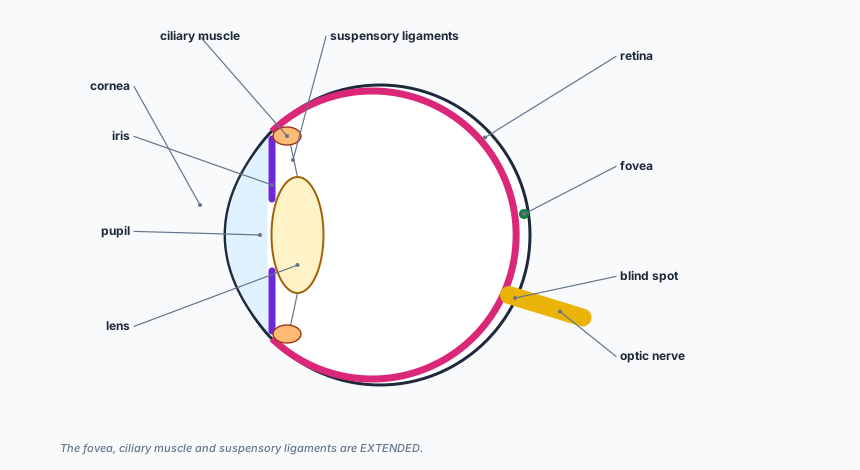
A horizontal section of the eye. At the front, the transparent cornea; behind it the coloured iris with the pupil, the gap in the middle; the lens held by suspensory ligaments attached to the ciliary muscle. The retina lines the back, with the fovea opposite the lens; the optic nerve leaves at the back, where the blind spot is.
The blind spot has no receptor cells, because the optic nerve leaves the eye there.
cornea: refracts light
iris: controls how much light enters the pupil
lens: focuses light on to the retina
retina: contains light receptors, some sensitive to light of different colours
optic nerve: carries impulses to the brain

The pupil reflex: in bright light the pupil becomes smaller, letting less light in and protecting the retina; in dim light it widens, letting more light in.
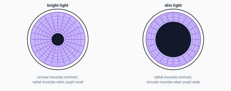
Two views of the iris from the front. In bright light the pupil is small: circular muscles contract and radial muscles relax. In dim light the pupil is wide: radial muscles contract and circular muscles relax.
EXTENDED The circular and radial muscles are antagonistic: when one contracts, the other relaxes.

EXTENDED Accommodation changes the shape of the lens to focus light from objects at different distances.
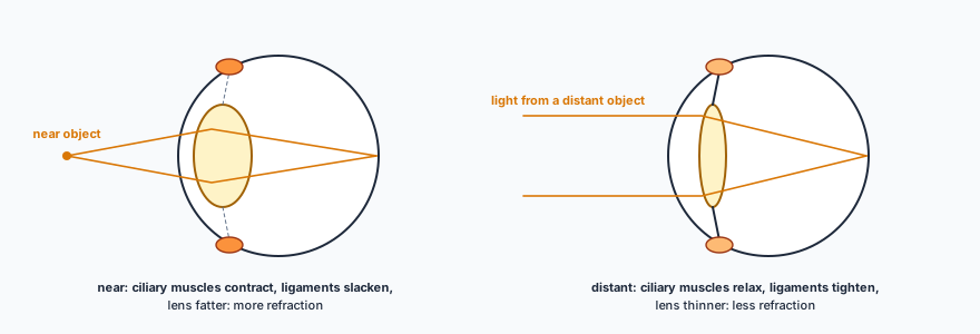
Two eyes. Focusing on a near object: diverging rays enter, the ciliary muscles are contracted, the suspensory ligaments slack and the lens fat, refracting light more to focus on the retina. Focusing on a distant object: parallel rays, ciliary muscles relaxed, ligaments tight, lens thin, refracting less.
EXTENDED Near: fat lens, more refraction. Distant: thin lens, less refraction.
  • EXTENDED Near object: the ciliary muscles contract, the suspensory ligaments slacken, the lens becomes fatter and refracts light more.
  • Distant object: the ciliary muscles relax, the suspensory ligaments tighten, pulling the lens thinner, so it refracts light less.
  • Rods are more sensitive to light, so they give night vision; they are found all over the retina except the fovea. Cones come in three kinds, absorbing different colours of light, for colour vision; they work only in bright light.
  • The fovea is the part of the retina where cones are packed most densely (no rods); it gives the sharpest, colour vision.

✏️Worked example

A person walks out of a dark room into bright sunlight. (a) Describe the change in the pupil and explain why it is useful. [2] (b) State the type of neurone that carries the impulse from the retina to the brain. [1] (c) EXTENDED Explain how the muscles of the iris bring about the change. [2] (d) EXTENDED The person then reads a label on a bottle held close up. Describe the changes in the eye. [3]

(a) The pupil becomes smaller, letting less light into the eye, which protects the retina from damage by bright light.

(b) A sensory neurone.

(c) The circular muscles contract and the radial muscles relax (they act antagonistically), making the pupil smaller.

(d) The ciliary muscles contract, the suspensory ligaments slacken, and the lens becomes fatter, refracting light more so the near object is focused on the retina.

Check it. Both answers (c) and (d) are pairs: one muscle set contracts while the other relaxes, and the lens shape follows from the ligaments.
“The iris gets smaller.” It is the pupil (the hole) that gets smaller; the iris covers more of the eye.

📝Practise

In the style of the multiple-choice and theory papers. EXTENDED marks Supplement content.

1. (Multiple choice.) What is the function of the optic nerve? A: refracts light. B: controls the size of the pupil. C: carries impulses to the brain. D: detects light.
C.
2. (Theory.) Name the two parts of the central nervous system. [2]
Brain; spinal cord.
3. (Theory.) A person steps on a sharp pin and lifts their foot before feeling pain. Name the receptor’s location, the effector, and explain why this reflex is useful. [3]
Receptor: in the skin of the foot. Effector: leg muscles. It is fast and automatic, so the foot is removed before much damage is done.
4. (Theory.) Compare the roles of the cornea and the lens. [2]
Both refract light. The cornea does most of the refraction; the lens changes shape to focus the light on to the retina.
5. (Theory.) State the function of the iris. [1]
It controls how much light enters the eye through the pupil.
6. (Theory.) EXTENDED Describe the events at a synapse. [4]
An impulse arrives and stimulates the release of neurotransmitter from vesicles into the synaptic gap; the neurotransmitter diffuses across the gap; it binds to receptor proteins on the next neurone; an impulse is stimulated in the next neurone.
7. (Theory.) EXTENDED Explain why impulses can only pass one way across a synapse. [2]
Neurotransmitter is released only from vesicles in the first neurone, and receptor proteins are only on the next neurone.
8. (Theory.) EXTENDED Explain why we cannot see colours well at night. [2]
In dim light only the rods work (they are more sensitive to light), and rods do not distinguish colours; the cones, which give colour vision, need bright light.

🔗Go deeper — other people’s work

These are external resources, not mine. If one stops working, tell me and everything above it on this page still stands.

  • Royal Society of Biology — measuring reaction time with a ruler drop
  • BBC Bitesize — the eye and focusing