Home › Learning Hub › IGCSE Biology › 2 Cells and size of specimens
Topic 2 · 2.1–2.2

Cell structure and the size of specimens

Core and Extended · Papers 1–6

🎯What you need to be able to do

  • Describe and compare plant, animal and bacterial cells, identify their structures in diagrams and images, and state the function of each structure.
  • State that new cells come from the division of existing cells, and give the functions of six specialised cells.
  • Use the terms cell, tissue, organ, organ system and organism.
  • Use magnification = image size ÷ actual size, in millimetres; convert between mm and µm EXTENDED.

📚The biology

Plant and animal cells
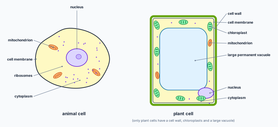
Labelled diagrams of an animal cell and a plant cell. The animal cell has a cell membrane, cytoplasm, a nucleus, mitochondria and ribosomes. The plant cell has all of these plus a thick cell wall, chloroplasts and a large permanent vacuole that fills most of the cell, with the nucleus pushed to the edge.
Both cells have a membrane, cytoplasm, nucleus, mitochondria and ribosomes. Only the plant cell has a cellulose cell wall, chloroplasts and a large permanent vacuole.
cell membrane: controls which substances enter and leave the cell
nucleus: contains the genetic material (chromosomes) that controls the cell’s activities
cytoplasm: where most of the cell’s chemical reactions take place
mitochondria: site of aerobic respiration, which releases energy
ribosomes: where proteins are made (protein synthesis)
cell wall (plants): made of cellulose; strengthens the cell and stops it bursting when it takes in water
chloroplasts (plants): contain chlorophyll, which absorbs light for photosynthesis
vacuole (plants): a large permanent space filled with cell sap; keeps the cell firm (turgid)

Not every plant cell has chloroplasts: a root hair cell is underground and has none. Cells with high energy demands, such as muscle cells and sperm, have many mitochondria.

Bacterial cells
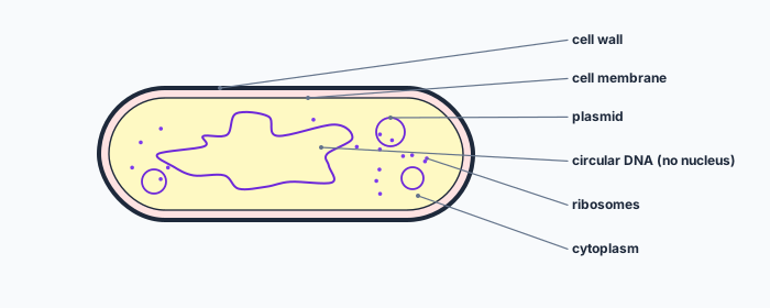
A rod-shaped bacterium with a cell wall on the outside, a cell membrane inside it, cytoplasm, scattered ribosomes, a loop of circular DNA lying free in the cytoplasm and three small rings called plasmids.
A bacterium has no nucleus: its main DNA is one circular loop in the cytoplasm. Plasmids are small extra rings of DNA.

Bacterial cells have a cell wall (not made of cellulose), a cell membrane, cytoplasm and ribosomes, but no nucleus and no mitochondria. Their genetic material is circular DNA, plus small circles called plasmids, which carry a few extra genes (for example for antibiotic resistance) and are used in genetic modification (topic 21).

Specialised cells and levels of organisation

New cells are produced by the division of existing cells. Many cells are then specialised: their structure suits one job.
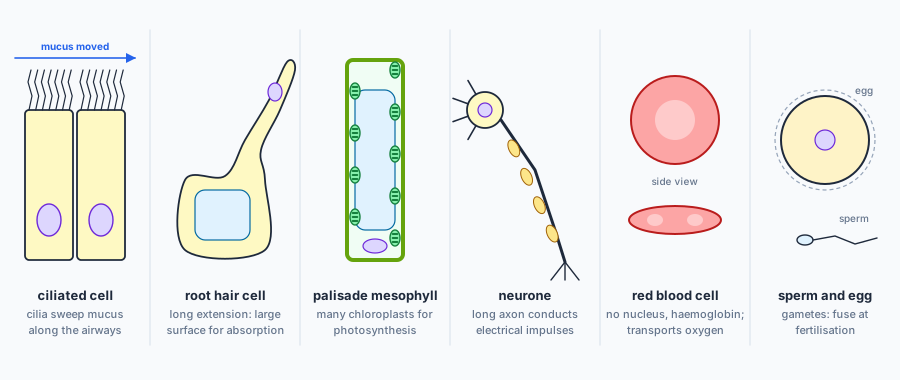
Six specialised cells. Ciliated cells with hair-like cilia that move mucus. A root hair cell with a long extension. A tall palisade mesophyll cell packed with chloroplasts. A neurone with a long axon. A red blood cell, a biconcave disc with no nucleus. A large egg cell and a small sperm cell with a tail.
The six specialised cells on the syllabus, each with the feature that fits its function.
ciliated cells: move mucus in the trachea and bronchi
root hair cells: absorption of water and mineral ions
palisade mesophyll cells: photosynthesis
neurones: conduction of electrical impulses
red blood cells: transport of oxygen
sperm and egg cells (gametes): reproduction

A tissue is a group of cells with similar structures working together to perform a shared function. An organ is a structure made of a group of tissues working together. An organ system is a group of organs with related functions working together, and an organism is a whole living thing.
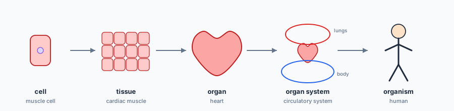
A sequence of five pictures joined by arrows: a single muscle cell, a block of cardiac muscle tissue, the heart as an organ, the heart with loops to the lungs and body as the circulatory system, and a whole human as the organism.
Cell → tissue → organ → organ system → organism. In a plant: palisade cell → palisade tissue → leaf → shoot system → plant.

Size of specimens

\[ \text{magnification} = \frac{\text{image size}}{\text{actual size}} \]

Measure the image with a ruler in millimetres, and use the same unit for both sizes: magnification has no units. Rearranged, actual size = image size ÷ magnification.

EXTENDED Cells are usually measured in micrometres (µm): 1 mm = 1000 µm. To convert mm to µm, multiply by 1000; to convert µm to mm, divide by 1000.

✏️Worked example
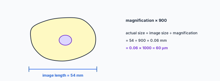
A student draws a human cheek cell seen through a microscope at a magnification of ×900. The drawing is 54 mm long. (a) Calculate the actual length of the cell in mm. [2] (b) EXTENDED Give your answer in micrometres. [1] (c) Name two structures a palisade mesophyll cell has that the cheek cell does not. [2]
A drawing of a cheek cell with its nucleus. A measurement bar under the drawing shows an image length of 54 millimetres. Beside it: magnification times 900; actual size = image size divided by magnification = 54 divided by 900 = 0.06 millimetres = 60 micrometres.

(a) actual size = image size ÷ magnification = \( 54 \div 900 = 0.06 \) mm.

(b) \( 0.06 \times 1000 = 60 \) µm.

(c) Any two: cell wall; chloroplasts; large permanent vacuole.

Check it. Human cells are roughly 10–100 µm across, so 60 µm is sensible. If you get a cell several millimetres long, you multiplied instead of dividing.
Mixing units. Put image size and actual size in the same unit before dividing. A 54 mm image divided by a 60 µm cell gives 0.9, not 900.

📝Practise

In the style of the multiple-choice and theory papers. EXTENDED marks Supplement content.

1. (Multiple choice.) Which structure is found in animal, plant and bacterial cells? A: nucleus. B: chloroplast. C: ribosome. D: large permanent vacuole.
C. Bacteria have no nucleus; animal cells have no chloroplasts or large vacuole.
2. (Theory.) State the function of mitochondria and suggest why a sperm cell contains many of them. [2]
Site of aerobic respiration, which releases energy. The sperm needs a lot of energy to swim (move its tail) to the egg.
3. (Theory.) A root hair cell has no chloroplasts. Explain why, and describe how its shape helps its function. [3]
It is underground, where there is no light for photosynthesis. Its long, thin extension gives a large surface area for absorbing water and mineral ions.
4. (Theory.) A photograph of a red blood cell is 21 mm across. The actual diameter is 0.007 mm. Calculate the magnification. [2]
\( 21 \div 0.007 = 3000 \), so ×3000.
5. (Theory.) Classify each as a cell, tissue, organ or organ system: (a) a leaf, (b) xylem, (c) a palisade mesophyll cell, (d) the digestive system. [2]
(a) organ, (b) tissue, (c) cell, (d) organ system.
6. (Theory.) State two ways in which a bacterial cell differs from a plant cell. [2]
Any two: no nucleus; circular DNA; plasmids; no chloroplasts; cell wall not made of cellulose; no large permanent vacuole; no mitochondria.
7. (Theory.) EXTENDED Convert (a) 0.25 mm to µm, (b) 7500 µm to mm. [2]
(a) \( 0.25 \times 1000 = 250 \) µm. (b) \( 7500 \div 1000 = 7.5 \) mm.
8. (Theory.) EXTENDED A cell 12 µm long is drawn at a magnification of ×2500. Calculate the length of the drawing in mm. [2]
image size = actual size × magnification = \( 12 \times 2500 = 30\,000 \) µm \( = 30 \) mm.

🔗Go deeper — other people’s work

These are external resources, not mine. If one stops working, tell me and everything above it on this page still stands.

  • Cells Alive! — interactive animal, plant and bacterial cell models
  • Royal Society of Biology — guide to using a light microscope and measuring cells