Circulation and the heart
🎯What you need to be able to do
- Describe the circulatory system; describe single and double circulation and explain the advantages of double circulation EXTENDED.
- Identify the structures of the heart; state that arteries carry blood away and veins return it; identify the valves EXTENDED.
- State how heart activity is monitored; investigate and explain the effect of physical activity on heart rate.
- Describe coronary heart disease, its risk factors, and the roles of diet and exercise in reducing it.
- Explain wall thickness, the septum and how the heart functions EXTENDED.
📚The biology
Circulatory systems
A circulatory system is a system of blood vessels with a pump (the heart) and valves to ensure one-way flow of blood.
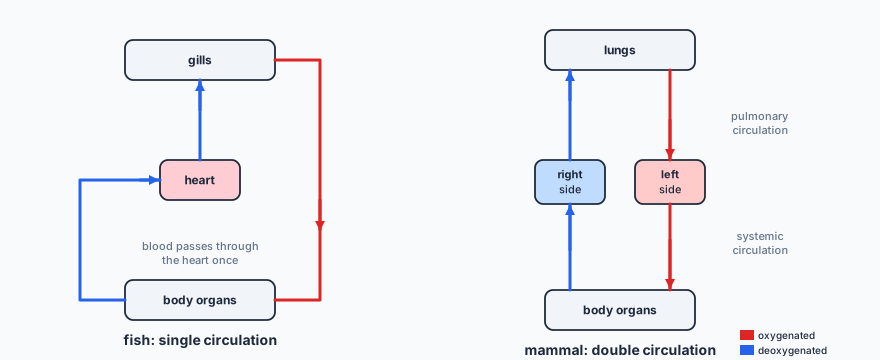
EXTENDED A fish has a single circulation: blood passes through the heart once per circuit, from heart to gills to body and back. A mammal has a double circulation: heart → lungs → heart → body → heart. The advantages: blood returning from the lungs is pumped again, so it reaches the body at high pressure and flows quickly, delivering oxygen and glucose fast enough for a high rate of respiration; the lungs receive blood at lower pressure, which does not damage their delicate capillaries.
The heart
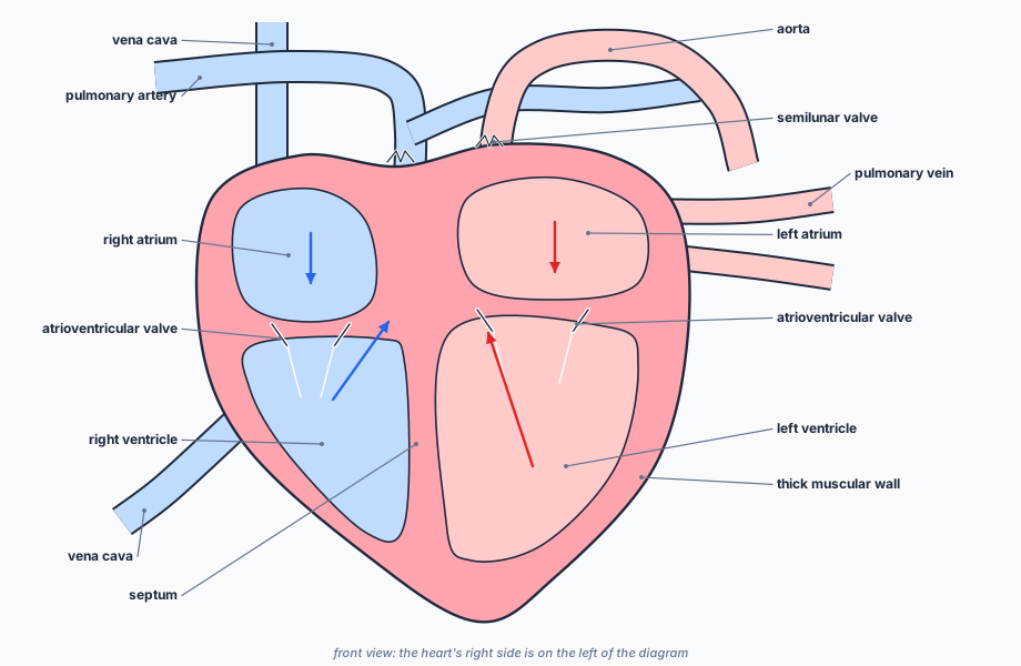
Blood is pumped away from the heart in arteries (aorta, pulmonary artery) and returns to it in veins (vena cava, pulmonary vein). The heart muscle itself is supplied with blood by the coronary arteries on its surface.
EXTENDED Features to explain:
- EXTENDED The left ventricle has a thicker muscle wall than the right, because it pumps blood at high pressure all round the body; the right pumps only to the nearby lungs.
- The atria have thinner walls than the ventricles: they only pump blood into the ventricles just below.
- The septum separates oxygenated and deoxygenated blood, so the body receives blood with as much oxygen as possible.
- Atrioventricular valves (between atria and ventricles) and semilunar valves (at the bases of the aorta and pulmonary artery) stop blood flowing backwards.
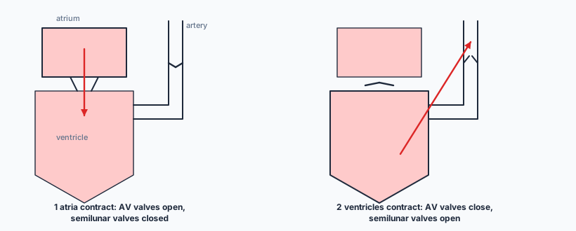
EXTENDED How the heart works: the atria contract, pushing blood through the open atrioventricular valves into the ventricles; then the ventricles contract, the atrioventricular valves close (so blood cannot return to the atria), and the semilunar valves open as blood is pushed into the arteries. When the ventricles relax, the semilunar valves close, stopping backflow from the arteries.
Monitoring the heart and exercise
Heart activity can be monitored by an ECG (electrocardiogram), by the pulse rate, and by listening to the sounds of the valves closing with a stethoscope. During physical activity the heart rate increases.
EXTENDED During exercise, muscles respire faster, needing more oxygen and glucose and producing more carbon dioxide. The heart beats faster (and more strongly) to deliver oxygen and glucose to the muscles faster and remove carbon dioxide faster.
Coronary heart disease
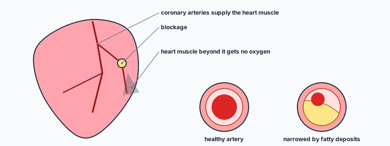
Coronary heart disease (CHD) is the blockage of coronary arteries, so heart muscle receives less oxygen and glucose, causing chest pain or a heart attack. Risk factors: a diet high in saturated fat and salt, lack of exercise, stress, smoking, genetic predisposition, age and sex (more common in males). A balanced diet low in saturated fat reduces fatty deposits; regular exercise strengthens the heart, lowers blood pressure and helps control body mass.
✏️Worked example
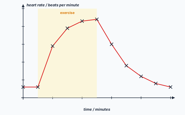
(a) 72 beats per minute.
(b) increase = \( 148 - 72 = 76 \); percentage increase = \( \tfrac{76}{72} \times 100 = 106\% \) (105.6%).
(c) Exercise stopped at 10 minutes; the rate was back to 72 at 20 minutes: 10 minutes.
(d) The muscles respire faster to release more energy for contraction. They need more oxygen and glucose, and produce more carbon dioxide, so the heart beats faster to deliver oxygen and glucose to the muscles and remove carbon dioxide more quickly.
📝Practise
In the style of the multiple-choice, theory and practical papers. EXTENDED marks Supplement content.
1. (Multiple choice.) Which blood vessel carries blood from the heart to the lungs? A: aorta. B: pulmonary artery. C: pulmonary vein. D: vena cava.
2. (Theory.) State three ways in which the activity of the heart can be monitored. [3]
3. (Theory.) State four risk factors for coronary heart disease, and one change to lifestyle that reduces the risk. [3]
4. (Practical.) Describe how to measure a person’s pulse rate accurately. [3]
5. (Theory.) EXTENDED Explain why the wall of the left ventricle is thicker than the wall of the right ventricle. [2]
6. (Theory.) EXTENDED A baby is born with a hole in the septum between the ventricles. Explain why the baby may lack energy and look slightly blue. [3]
7. (Theory.) EXTENDED Describe what happens to the valves when the ventricles contract. [2]
8. (Theory.) EXTENDED State one advantage of a double circulation over a single circulation. [1]
🔗Go deeper — other people’s work
These are external resources, not mine. If one stops working, tell me and everything above it on this page still stands.
- British Heart Foundation — how the heart works, with animations
- Royal Society of Biology — investigating the effect of exercise on pulse rate